CT Scans and 3D Computer-Assisted Virtual Implant Diagnostics
Dental cone beam imaging, a type of CT scanning, is used by Portland Perio Implant Center to obtain accurate diagnostic information. Computer Tomography (CT) is an imaging method that uses computerized technology to convert 2-dimensional images into a 3- dimensional (3D) picture. These pictures can provide renderings of both soft and hard tissues in the body which then can be examined in detail using computer software.
Dental cone beam imaging provides the doctor with more accurate information and 10 times less radiation to the patient than the machines traditionally used in medicine. What this means to patients who have chosen to replace missing teeth, is that now the doctor can examine, diagnose and plan treatment with a level of precision not possible in the past. This more exact approach using dental cone beam imaging means fewer complications, less invasive therapy, and quicker healing times for the patient.
Specifically, the 3D representation of the jaw seen on the CT scan enables the implant treatment team (the restorative dentist, periodontal surgeon and dental lab technician) to see the patient anatomy available for dental implants. The treatment team is able to work with an exact replica of the patient’s jawbone and gum tissue when choosing the size, location and angulation of the implant. Any procedures required to prepare the site for implants can be performed “virtually” on the computer using the dental cone beam tomography. This software is accurate within a tenth of a millimeter and shows the implant in its ideal position. Precise implant placement is critical in order to provide the patient the most esthetic and functional form of tooth replacement.
In some cases, a surgical guide based on the cone beam imaging result is fabricated by the dental lab prior to treatment. This customized guide allows the surgeon to perform the procedure without any incisions at all. Less invasive treatment means more comfort for the patient and quicker healing times.
The Academy of Osseointegration now considers dental cone beam technology “the standard of care” for implant procedures. In fact, The American Board of Periodontology and American Academy of Oral and Maxillofacial Surgeons advises the use of exact imaging and surgical guides for dental implant placement. Unfortunately, due to the great expense and training required to provide this service to patients, not all offices currently utilize CT scanning technology in their offices.
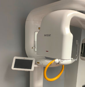
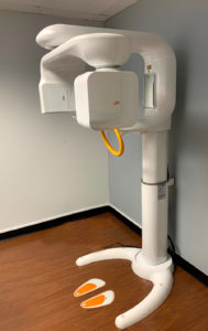
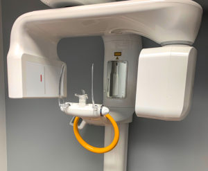
Our CT scanner is a Rayscan Alpha Plus, with cutting edge detectors and x-ray technology. We can scan precisely where we need to, with the added benefit of low-dose capability for safer scanning for our patients. It provides advanced 3D treatment tools for implants and restorations, oral and maxillofacial surgery, TMJ and sinuses with high definition 3D diagnostic images for ultimate treatment efficiency.
Once a CT Scan is captured, a 3-dimensional view is created and implant placement is virtually planned with very high accuracy. All can be done within minutes of the scan. The data from our case planning is then replicated with great precision in the patient on the day of implant placement.


